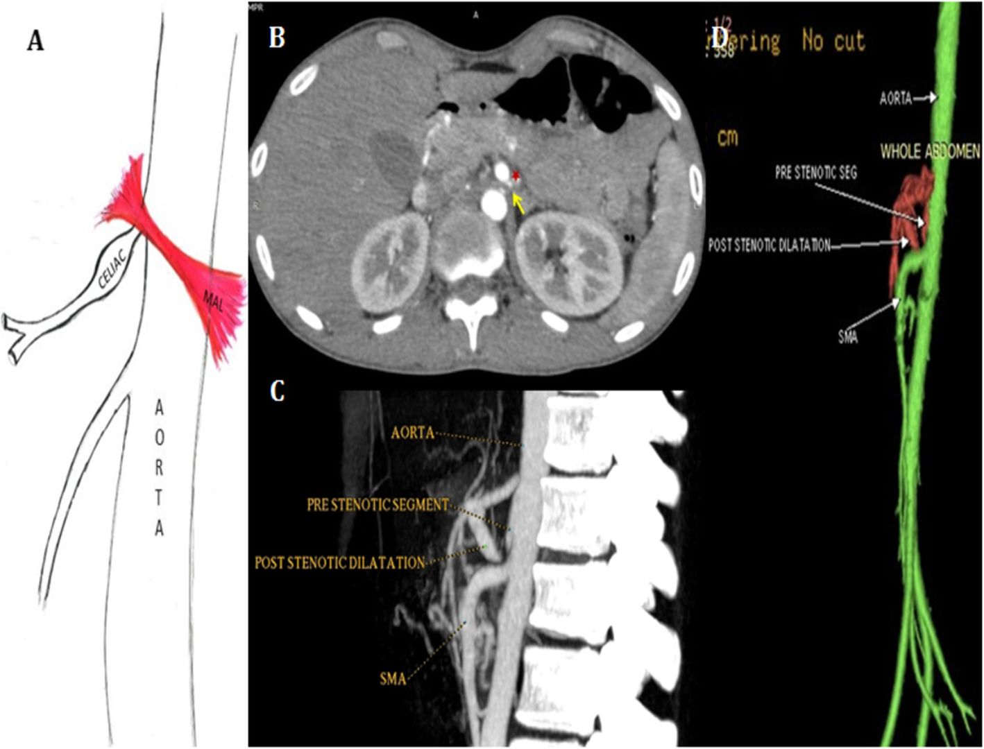
Fig. 1

A 37-year-old case with median arcuate ligament syndrome. (a) shows a hand diagram of celiac trunk origin illustrating the compression of the origin of the coeliac trunk by the median arcuate ligament with post stenotic dilatation. Axial view of the contrast-enhanced CT study of the abdomen in arterial phase (b) shows the median arcuate ligament (arrow) compressing the origin of the coeliac trunk with post stenotic dilatation (asterisk). Sagittal reformatted MIP (c) and volume-rendered (d) images show the median arcuate ligament compressing the origin of the coeliac trunk with post stenotic dilatation. The hand diagram is self-drawn by the author