- Research
- Open access
- Published:
The role of MR venography with time-resolved imaging in diagnosis of pelvic congestion syndrome
Egyptian Journal of Radiology and Nuclear Medicine volume 53, Article number: 19 (2022)
Abstract
Background
Pelvic congestion syndrome (PCS) represents a diagnostic challenge due to its variable clinical presentation, complex anatomy, and pathophysiology. Accurate delineation of the venous anatomy, detection of venous reflux or obstruction, its extent will enable interventional radiologists to successfully treat such patients and to avoid recurrence. Magnetic resonance imaging (MRI) allows a noninvasive examination of the anatomy and flow inside the pelvic veins in addition to its excellent soft-tissue contrast allowing evaluation of the pelvic organs. Our study is aiming to investigate the role and accuracy of MR venography with time-resolved imaging (TR-MRV) as a diagnostic tool for pretreatment planning of PCS.
Results
Our study included 25 female patients with mean age 48 ± 12.34, who were referred to the radiology department in the period from April/2019 to April/2020 with clinical and ultrasound features suggesting PCS. TR-MRV was performed and interpreted in a blind fashion evaluating the vascular anatomy, venous dilatation, and reflux. The results were compared to conventional venography as a reference. The sensitivity, specificity, and accuracy of TR-MRV in the detection of ovarian vein reflux were 87%, 80%, and 84%, respectively, versus 75%, 53%, and 72% in internal iliac vein reflux and 92%, 69%, and 64% for pelvic venous plexus reflux. Demonstration of the venous anatomy was excellent in 68% of the patients and was sufficient in 32%. Ovarian vein dilatation was detected in 16 patients by venography and in 10 patients by TR-MRV. The weighted k-values (Cohen's Kappa coefficient statistics) indicated excellent agreement between the two observers for identifying all the refluxing veins by TRI in each patient (k = 0.78).
Conclusion
MRI with TR imaging has shown high diagnostic accuracy when compared to conventional venography in evaluating pelvic congestion syndrome before endovascular treatment and thus facilitating treatment planning.
Background
Pelvic congestion syndrome (PCS) is a complex, underdiagnosed cause of chronic pelvic pain in female patients. Up to 38 out of 1,000 women annually present in primary care with intermittent or constant pain in the lower abdomen or pelvis, a rate comparable with that of Asthma and lower back pain (37 and 41 in 1000, respectively) [1]. However, pelvic pain may be caused by many other entities including gynecological, urological, gastrointestinal, and musculoskeletal [2]. Therefore, imaging is critical in the evaluation of PCS, to differentiate it from other conditions and to detect its cause [3].
Multiple etiologies may be responsible for Pelvic Congestion Syndrome. PCS caused by incompetent gonadal vein valves is termed pelvic venous insufficiency (PVI) [4]. Secondary causes include retro-aortic left renal vein, meso-aortic compression of the left renal vein (nutcracker syndrome), May–Thurner syndrome, IVC obstruction, vascular malformations, or portal hypertension. Hormonal factors/reproductive age and multiple pregnancies are also widely acknowledged risk factors [5].
Most women present with non-cyclic, intermittent, constant pelvic pain for more than 6 months. Other symptoms include dyspareunia, dysmenorrhea, and urinary urgency. The clinical examination may reveal varicose veins of the vulva, perineum, buttocks, and lower extremities [2]. However, pelvic venous engorgement and gonadal vein reflux can be seen in patients without pelvic pain. Thus, the diagnosis of pelvic congestion syndrome is made based on the patient's symptoms, clinical examination, and imaging studies.
Angioembolization of the ovarian veins and pelvic varices is the treatment of choice for PVI. Treatment failure is explained by the complex anatomy of the pelvic veins, which show a wide variation in terms of trunks, venous valves, duplications, and crossover connections. Furthermore, reflux often affects more than one pelvic vein which makes it difficult to identify and treat all patterns of reflux leading to the development of alternative reflux pathways and recurrence after treatment [6].
Venography is regarded as the "gold standard" for diagnostic studies which can show retrograde flow in the ovarian and pelvic veins, incompetent pelvic veins, congestion of flow in the ovarian, pelvic, vulvovaginal, thigh veins [7]. Patient discomfort due to its invasive nature, use of ionizing radiation, costs is major drawbacks [8]. The diagnosis of PCS has usually been suggested by duplex ultrasound (US), but ultrasound imaging does not readily show the gonadal veins in addition to its limited sensitivity for the identification of structural causes of PCS and other conditions that cause pelvic pain (e.g., endometriosis) [9].
Magnetic resonance imaging (MRI) can demonstrate anatomic findings such as dilated ovarian veins, the presence of pelvic, perineal, vulval/labial, thigh varices. Secondary causes of PVI can also be detected with high accuracy because of its multiplanar imaging capability and excellent soft-tissue contrast [10]. Magnetic resonance venography (MRV) with time-resolved imaging (TRI) is a quick and noninvasive technique that allows evaluation of the dynamic blood flow pattern and is particularly useful for abdominal venous imaging in free-breathing patients and when the presence and direction of flow are critical to the diagnosis [11]. Accurate anatomic detail and flow information are essential to provide a detailed roadmap for the embolization procedure, detecting variant anatomy, and ruling out non-PVI etiologies. This study aims to evaluate the role and accuracy of TR-MRV in the diagnostic work-up of pelvic congestion syndrome before endovascular treatment.
Methods
Patients
This prospective study included 25 women with a mean age of 48.67 ± 12.34 (range 34–55) who were referred to the radiology department with clinical and US features suggesting PCS, in the period from April/2019 to April/2020. At the time of patient hospital admission, comorbidities, as well as standard physical examination findings, were recorded.
Clinical examination
The clinical evaluation included a history of congestion symptoms and a physical examination of the pelvic region and the lower limbs to demonstrate signs of venous incompetence or obstruction. This included four clinical presentations (a) chronic pelvic pain of at least 6 months; (b) venous claudication due to iliac venous obstruction; (c) left flank or abdominal pain and hematuria due to left renal vein compression; and (d) symptomatic lower extremity varicosities in either atypical (vulva, medial, and posterior thigh, sciatic nerve) or typical saphenous distributions.
Any patient with general contra-indications to MRI as the presence of any paramagnetic substance as pacemakers, or in severely ill patients or those with claustrophobia, arrhythmic patients (that were managed by pacemakers) were excluded from the study that has a pacemaker. The study was conducted after approval of the institutional Ethical Committee, and informed written consent was obtained from each participant.
MRI, and TR-MRV
The MRI examinations were performed with a 1.5 T imaging unit (Magnetom Sempra, Siemens, Erlangen, Germany) with the use of a body phased-array coil. Preliminary sequences covering the abdomen and pelvis from the upper pole of the left kidney to the proximal thighs were performed for all patients. These included axial, coronal, and sagittal T2-weighted turbo spin-echo (T2_TSE) images and axial T1-weighted fast low angle shot (FLASH) images to examine the pelvic organs and detect dilatation of the pelvic veins. The T2_TSE sequence was performed with the following parameters: repetition time (TR)/echo time (TE) 3570/100 ms; field of view of 210 mm; flip angle 150 and slice thickness of 4 mm. T1_FLASH sequence was performed with the following parameters: TR/TE of 185/5 ms; field of view 360 mm; flip angle 70 and slice thickness 6 mm.
MRV with TRI was then performed for the detection of venous reflux. The acquisition parameters of TR-MRA were the following: TR/TE of 5.5/1.5 ms, flip angle 25, slices/slab 70, slice thickness 1 mm, and field of view 360 mm. Four phases (arterial, late arterial, venous, and late venous) were performed in the coronal plane during shallow breathing for 2 min after iv injection of 0.1 mmol/kg bodyweight of contrast medium gadopentetate dimeglumine (Magnevist, Schering, Berlin, Germany), at a rate of 2 ml/s which was followed by a 20 ml saline flush. To achieve maximum contrast signal in the veins, the transit time of the contrast medium was determined with the use of a bolus tracking technique initiated by abdominal aortic enhancement at the renal artery level. The acquisition time per phase was 20 s, and the intervals between phases were 5 s. The post-enhanced MR images for each phase were subtracted from the pre-contrast MR image and used to generate MRV. The images are then post-processed using maximum intensity projection (MIP) algorithms.
Finally, a post-contrast axial 3D fat-suppressed T1-weighted (T1_VIBE) sequence covering the abdomen and pelvis was performed with the following parameters: TR/TE of 5.82/2.35 ms; field of view 320 mm; flip angle of 10 and slice thickness of 3 mm.
Venography
Venography was performed using the Artiszee-Siemens Healthcare Angiography system. With the patient in a supine position, venographic access was obtained with the Seldinger technique via the right femoral vein or the right internal jugular vein. Catheterization of the inferior vena cava was done using a 5-French Cobra catheter (Cook, Bloomington, IN), and bilateral venography of the common iliac vein, external iliac vein, and a selective internal iliac vein was performed to evaluate for reflux in the internal iliac veins and narrowing of the left common iliac vein in May–Thurner syndrome. Venography of the left renal vein was then performed to detect nutcracker syndrome or reflux into the left ovarian vein. If reflux is seen in the ovarian vein, the catheter was positioned in the proximal part of the vein and selective venography was performed to detect reflux into the visceral venous plexus and bridging arcuate uterine veins to the contralateral side. Finally, a contrast medium is injected into the inferior vena cava to detect incompetence of the right ovarian vein and selective right ovarian vein venography was performed if needed.
At the end of the examination, patients were observed for about 4 h and then discharged home.
Image analysis
MRI and MRV with TRI were interpreted first in a prospective and blinded manner by two radiologists with 15 and 20 years of experience, and diagnosis was made in consensus. Conventional venography was used as the standard of reference. For the analysis, the venous system was divided into the following segments: the common and internal iliac veins, the ovarian veins, and the pelvic plexus.
Demonstration of the venous anatomy on TRI was assessed as either inadequate (impossible to definitively determine a treatment plan), sufficient (intermediate image quality but sufficient for treatment planning), or excellent anatomic visualization.
The diagnostic criteria for pelvic venous incompetence on MRV with TRI were the retrograde caudal flow of contrast material, dilated para-uterine varices, the presence of an arcuate vein crossing the midline, heterogeneous or T2-hyperintensity due to slow flow, vulvar and/or thigh varices, polycystic ovarian configuration and the absence of an obstructing mass or structural obstruction.
The diagnostic criteria for pelvic venous incompetence on venography were dilated gonadal, uterine, and utero-ovarian arcade veins > 5 mm in diameter, retrograde caudal flow in the ovarian vein (unilateral or bilateral), reflux of contrast material across the midline to the contralateral side through the utero-ovarian arcade, retrograde filling of the principal tributaries of the IIV (gluteal, sciatic, obturator vein) and stagnation of contrast material in pelvic veins [4]. May–Thurner Syndrome was diagnosed by compression of the left common iliac vein by the right common iliac artery.
Statistical analysis
All statistical tests were performed using IBM-SPSS 23.0 (IBM-SPSS Inc., Chicago, IL, USA). Continuous data were expressed in mean and standard deviation while categorical data were expressed in count and percentage. Weighted k statistics were calculated to assess the interobserver agreement for correctly identifying all the refluxing veins by TRI in each patient. The level of agreement was defined as follows: k-values of 0.00–0.40 indicated poor agreement, k-values of 0.41–0.75 indicated good agreement, and k-values of 0.76–1.00 represented excellent agreement.
Results
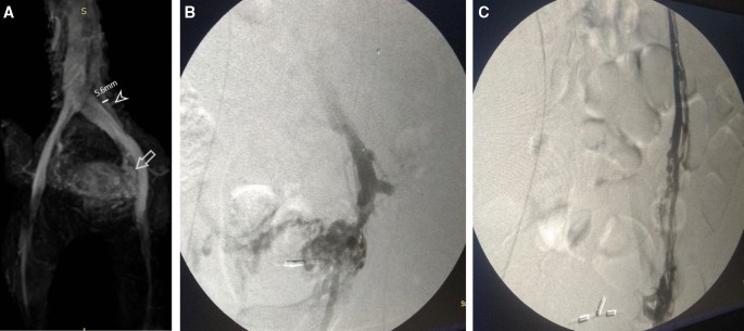
The 25 women included in this study presented with chronic pelvic pain. Other symptoms included pelvic heaviness, labial varicosities, lower limb varicosities, dyspareunia, and venous claudication (Table 1). Venography revealed that 22 of the 25 women with clinical signs and symptoms of PCS had one or more incompetent pelvic veins while TRI detected incompetence in 20 patients. One patient was diagnosed by both modalities with PVI secondary to May–Thurner syndrome (Fig. 1).
A Case 1 MRV 3D image that shows dilated left ovarian vein reaching (5.6 mm), with dilated left pelvic varices. B Case 1 conventional direct venography: shows dilated distal part of the left ovarian vein and pelvic plexus. C Case 1 conventional venography: same case, but contrast administration was done at the proximal part of the left ovarian vein, to show the whole vein diameters and week points
Demonstration of the venous anatomy on MRV with TRI was excellent in 17 (68%) patients and sufficient in 8 (32%) patients with no inadequate examinations. Ovarian vein dilatation was detected in 16 patients by venography and in 10 patients by TRI with one false-positive result. MRV also did not detect dilatation of the internal iliac veins in 2 patients and the pelvic plexus in 2 patients.
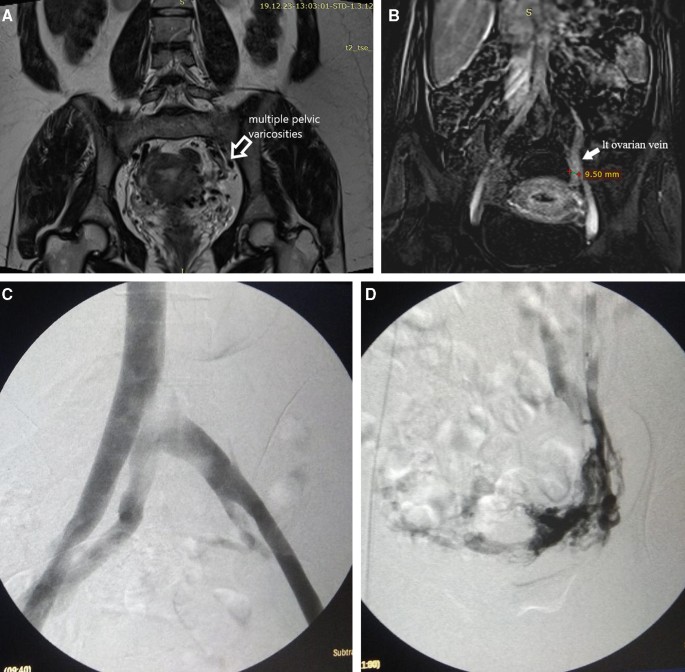
Reflux was detected by venography in 15 (60%) ovarian veins, 4 (16%) internal iliac veins, and 12 (48%) pelvic plexuses, while TRI detected 13 (52%), 3 (12%), and 9 (36%), respectively (Table 2). MRV with TRI had 87% sensitivity and 80% specificity for detection of ovarian venous incompetence in patients with PCS with an overall accuracy of 84% and an area under the curve of 0.83 (Table 3 and Figs. 2, 3). For detection of iliac venous incompetence, TRI had 75% sensitivity and 53% specificity with an overall accuracy of 72% and an area under the curve of 0.76 (Table 3). Regarding pelvic venous plexus incompetence, it had 92% sensitivity and 69% specificity with an overall accuracy of 72% and the area curve was 0.80 (Table 3).
A Case 2 MRI. (T2 conventional image) shows multiple dilated signal void pelvic varicosities. B Case 2 MRV (time-resolved image after contrast administration that shows dilated left ovarian vein reaching (9.5 mm). C Case 2 conventional venography shows dilated left ovarian vein secondary to compression by the right iliac artery (May–Turner Syndrome). D Case 2 conventional venography (shows dilated left ovarian vein and left-sided pelvic plexus secondary to May–Turner Syndrome, As Described)
A1 Case 3 MRV (time-resolved coronal image after contrast administration with dilated pelvic varicosities). A2 Case 3 MRV (Time-resolved axial image after contrast administration that shows multiple dilated pelvic varicosities). B1 Case 3 conventional venography (show dilated left ovarian vein along its whole length with multiple week points). B2 Case 3 conventional venography (selective catheterization of the proximal part of the left ovarian vein that shows its dilatation with multiple dilated pelvic varicosities
The weighted k-values indicated excellent agreement between the two observers for identifying all the refluxing veins by TRI in each patient (k = 0.78).
Discussion
Chronic pelvic pain is a common health problem among women and is defined as noncyclic pelvic pain of more than 6 months duration. The condition is potentially debilitating, and diagnosing its cause can be quite challenging for clinicians. PCS is one of the causes of chronic pelvic pain which is often overlooked in the differential diagnosis due to its non-specific clinical presentation. The various etiologies of PCS are responsible for its diverse symptoms. PCS may be due to incompetent gonadal vein valves and is termed pelvic venous insufficiency or secondary to structural causes such as left renal vein compression with an incompetent gonadal vein valve (nutcracker syndrome) or iliac vein compression (May–Thurner configuration) with reflux into the ipsilateral internal iliac vein [3].
Ovarian and pelvic vein embolization is now the standard method of treatment of PCS using embolization coils, sclerosants, or a combination of both [4]. Success rates of up to 80% have been reported after a follow-up of 5 years [3]. However, to achieve such a high success rate, multiple essential points must be considered before treatment. First, anatomical venous variations including internal iliac veins that drain into the contralateral common iliac vein, duplicated IVCs, reverse-angle renal veins with alternative left gonadal vein drainage, and drainage of the right gonadal vein into the right renal vein may alter the approach to treatment. Furthermore, recognizing the blood flow dynamics in the pelvic veins is also essential for the diagnostic work-up of PCS [12]. The ovarian veins typically have multiple tributaries that generally collateralize into the utero-ovarian venous arcade with contralateral reflux of contrast medium across the midline. There is often opacification of vulvar or thigh varices and stagnation of contrast medium in pelvic veins. Incomplete embolization of the ovarian vein tributaries and associated pelvic collateral vessels may be a source of clinical failure [4]. Therefore, an imaging modality that allows accurate delineation of the complex venous anatomy and the reflux patterns of the pelvic veins is mandatory for treatment planning and success (Table 4).
The diagnosis of PCS is made through a combination of the patient’s symptoms, clinical and radiological examination. The gold standard test for diagnosing PCS is Venography. However, due to the invasive nature of venography and exposure to ionizing radiation, it is reasonable to limit its use to those patients with a high suspicion of PCS with the intent to treat it. This makes it necessary to have a reliable, noninvasive imaging modality that is suitable for screening and can yield consistent results with the patient’s clinical presentation [13].
Ultrasonography with color Doppler allows a dynamic examination of flow in the pelvic veins. Dilated peri-uterine and peri-ovarian veins can be identified in Pelvic Congestion Syndrome. It has the advantage of using the Valsalva maneuver or examining the patients in an upright position to detect reflux [14]. However, sonography does not readily show the ovarian veins and has limited sensitivity for obstructive causes of PCS and other conditions that cause pelvic pain (e.g., endometriosis) [9].
The diagnosis of PCS on static CT and MRI (without TRI) is based mainly on dilatation of the peri-uterine and peri-ovarian venous plexus and ovarian veins. In this study, ovarian vein dilatation was recorded in 10 (40%) patients by MRI as compared to 16 (64%) patients diagnosed by venography with only one false-positive result. Yang et al. [11] suggested that the discrepancy in the diameter of the left ovarian vein between TRI and venography may be due to the pressure effect on the left ovarian vein by selective injection of contrast medium into the left renal vein and the radiographic magnification of conventional venography. However, ovarian vein diameter is a poor predictor of gonadal vein reflux, and dilated pelvic veins can often be incidentally found in asymptomatic women. Therefore, the consensus statement from the Society of Interventional Radiology states that the absolute diameter of the veins should not preclude treatment of PVI in the presence of other findings [15].
In the current study, MRV with TRI provided an excellent demonstration of the venous anatomy in 17 (68%) patients and sufficient demonstration for treatment planning in 8 (32%) patients. The results of Asciutto et al. also revealed that visualization of venous anatomy was excellent or more than sufficient for treatment planning in all cases on MRV. In addition, Chennur et al. [16] stated that TRI provides accurate information about arterial anatomy and flow characteristics and thus, can detect any incidental AV malformations. This indicates that TRI is well-suited for the morphologic assessment of the pelvic and ovarian veins.
MRV with TRI enables the visualization of blood flow dynamics in addition to the excellent soft-tissue contrast and multiplanar capabilities of MRI allowing the detection of various associated pathologies when compared with conventional angiography. [17]. Furthermore, incompetence is frequently diagnosed in more than one pelvic vein, which highlights the importance of examining all the pelvic veins to achieve adequate treatment and avoid recurrence [6].
Our results revealed that the sensitivity and specificity of MRV with TRI for the detection of reflux were 87% and 80% for the ovarian veins, 75% and 53% for the internal iliac veins, and finally 92% and 69% for the pelvic venous plexus, respectively. The results of Asciutto et al. [18] also revealed that MRV is highly sensitive for insufficiency in pelvic plexus, ovarian or internal iliac veins. They reported 100% sensitivity when examining the internal iliac vein, but the specificity was low, which resulted in a high prevalence of false-positive results. Kim et al. [17] concluded that TRI is a useful imaging technique for the detection of ovarian vein reflux and that it could become the gold standard for the evaluation of pelvic venous congestion and chronic pelvic pain. Chennur et al. attributed the lower sensitivity of TRI in some cases due to early subtle reflux which can be missed on TRI due to supine patient positioning; unlike venography which allows table tilt and Valsalva maneuver [16]. However, other authors believe that TRI increases the sensitivity and specificity of MRI by demonstrating the true flow dynamics due to injection of contrast through peripheral intravenous access, allowing a more physiologic description of the flow characteristics in contrast to conventional venography where the flow dynamics can be altered due to the pressure of direct injection which can result in a false-positive diagnosis [17].
There was an excellent agreement between the two observers in identifying all the refluxing veins in each patient using TRI (k = 0.78). This agrees with the results of Yang et al. [11] who found excellent agreement between the two observers for grading ovarian venous reflux on TR-MRI (k = 0.894). All these factors indicate that MRI with TRI can be used as a reliable screening method for the initial evaluation of patients with suspected pelvic venous congestion [17].
The limitation of this study was that MRV may underestimate venous disease because conventional cross-sectional imaging studies are generally performed in the supine position in which ovarian and pelvic varices may not be as prominent. Another limitation is the relatively small number of patients included in the study. We recommend that a multicenter study be done with a large sample size to confirm the role of MRV in the diagnosis of PCS. Furthermore, a comparative study between different imaging modalities in the evaluation of women with suspected PCS as MRV, Duplex ultrasonography, and computed tomography would be of great value.
Conclusion
Although venography is the gold standard for the diagnosis of PCS, in the current study, MRV with TRI has shown to be a reliable noninvasive method in the evaluation of all aspects of PCS that are required for endovascular treatment planning with high diagnostic accuracy.
Availability of data and materials
The data that support the findings of this study are available from Radiology department-Assiut University, but there are restrictions apply to the availability of data, which are used under license for this study, and so were not publicly available. Data were available from authors upon request with permission of the head of the Radiology department- Assiut University.
Abbreviations
- PCS:
-
Pelvic congestion syndrome
- PVI:
-
Pelvic venous insufficiency
- MRI:
-
Magnetic resonance imaging
- TR-MRV:
-
Time-resolved. Magnetic resonance venography
- TRI:
-
Time-resolved imaging
References
Champaneria R, Shah L, Moss J, Gupta JK, Birch J, Middleton LJ et al (2016) The relationship between pelvic vein incompetence and chronic pelvic pain in women: systematic reviews of diagnosis and treatment effectiveness. Health Technol Assess 20(5)
Phillips D, Deipolyi AR, Hesketh RL, Midia M, Oklu R (2014) Pelvic congestion syndrome: etiology of pain, diagnosis, and clinical management. J Vasc Interv Radiol 25(5)
Kim HS et al (2006) Embolotherapy for pelvic congestion syndrome: long-term results. J Vasc Interv Radiol 17(2):289–297
Black CM, Thorpe K, Venrbux A et al (2010) Research reporting standards for endovascular treatment of pelvic venous insufficiency. J Vasc Interv Radiol 21(6):796–803
Borghi C, Dell’ Atti L (2016) Pelvic congestion syndrome: the current state of the literature. Arch Gynecol Obstet 293(2)
Asciutto G (2012) Pelvic vein incompetence: a review of diagnosis and treatment. Phlebolymphology 19(2):84–90
Geier B, Barbera L, Mumme A et al (2007) Reflux patterns in the ovarian and hypogastric veins in patients with varicose veins and signs of pelvic venous incompetence. Chir Ital 2007:59
Nascimento AB, Mitchell DG, Holland G (2002) Ovarian veins: magnetic resonance imaging findings in an asymptomatic population. J Magn Resonan Imaging 15(5):551–556
Bookwalter CA, VanBuren WM, Neisen MJ, Bjarnason H (2019) Imaging appearance and nonsurgical management of pelvic venous congestion syndrome. Radiographics 2019(39):596–608. https://doi.org/10.1148/rg.2019180159)
Watanabe Y et al (2000) Dynamic subtraction contrast-enhanced MR angiography: technique, clinical applications, and pitfalls. Radiographics 20(1):135–152
Yang DM, Kim HC, Nam DH, Jahng GH, Huh CY, Lim JW (2012) Time-resolved MR angiography for detecting and grading ovarian venous reflux: comparison with conventional venography. Br J Radiol 85(1014):e117–e122. https://doi.org/10.1259/bjr/79155839]
Beckett D, Dos Santos SJ, Dabbs EB, Shiangoli I, Price BA, Whiteley MS (2018) Anatomical abnormalities of the pelvic venous system and their implications for endovascular management of pelvic venous reflux. Phlebology 33(8):567–574. https://doi.org/10.1177/0268355517735727
Kelly DM, Sanford D, Stoughton J (2016) A study comparing the results of duplex ultrasound and magnetic resonance venography to diagnose pelvic vein congestion in conjunction with a compression syndrome. J Vasc Ultrasound 40(1):14–19
Park SJ, Lim JW, Ko YT, Lee DH, Yoon Y, Oh JH et al (2004) Diagnosis of pelvic congestion syndrome using transabdominal and transvaginal sonography. Am J Roentgenol 2004:182
Dos Santos SJ, Holdstock JM, Harrison CC, Lopez AJ, Whiteley MS (2015) Ovarian vein diameter cannot be used as an indicator of ovarian venous reflux. Eur J Vasc Endovasc Surg 49(1):90–94
Chennur VS, Nzekwu EV, Bhayana D, Raber EL, Wong JK (2019) MR venography using time-resolved imaging in the interventional management of pelvic venous insufficiency. Abdom Radiol 2019(44):2301–2307. https://doi.org/10.1007/s00261-019-01965-w
Kim CY, Miller MJ, Merkle EM (2009) Time-resolved MR angiography as a useful sequence for assessment of ovarian vein reflux. AJR Am J Roentgenol 2009(193):W458–W463
Asciutto G, Mumme A, MarpeB KO, Asciutto KC, Geier B (2008) MR venography in the detection of pelvic venous congestion. Eur J Vasc Endovasc Surg 36:491–496. https://doi.org/10.1016/j.ejvs.2008.06.024
Acknowledgements
The authors thank all the study participants for their patience and support.
Funding
No.
Author information
Authors and Affiliations
Contributions
N. M. A. Suggest and develop the research idea, reviewing the literature. M. A. S. was responsible for data collection and analysis, perform statistical analysis, write and revise the manuscript, prepare cases, perform required measurements, and prepare figures and tables. N. M. A, and M.A. S. were responsible for reporting the cases. All authors have a major contribution in preparing and editing the manuscript. All authors read and approved the final manuscript.
Corresponding author
Ethics declarations
Ethics approval and consent to participate
This study had approval from Egypt, Assiut University, Faculty of Medicine Research Ethics Committee (IRB Number 17100315).
Informed consents
Not applicable, verbal consent is mandatory as the procedure does not carry any significant risk for the patient.
Consent for publication
All patients included in this research gave written informed consent to publish the data contained within this study.
Competing interests
Authors declare that they had no competing interests.
Additional information
Publisher's Note
Springer Nature remains neutral with regard to jurisdictional claims in published maps and institutional affiliations.
Rights and permissions
Open Access This article is licensed under a Creative Commons Attribution 4.0 International License, which permits use, sharing, adaptation, distribution and reproduction in any medium or format, as long as you give appropriate credit to the original author(s) and the source, provide a link to the Creative Commons licence, and indicate if changes were made. The images or other third party material in this article are included in the article's Creative Commons licence, unless indicated otherwise in a credit line to the material. If material is not included in the article's Creative Commons licence and your intended use is not permitted by statutory regulation or exceeds the permitted use, you will need to obtain permission directly from the copyright holder. To view a copy of this licence, visit http://creativecommons.org/licenses/by/4.0/.
About this article
Cite this article
Attia, N.M., Sayed, M.A., Galal Mohamed, H.E. et al. The role of MR venography with time-resolved imaging in diagnosis of pelvic congestion syndrome. Egypt J Radiol Nucl Med 53, 19 (2022). https://doi.org/10.1186/s43055-021-00687-8
Received:
Accepted:
Published:
DOI: https://doi.org/10.1186/s43055-021-00687-8


